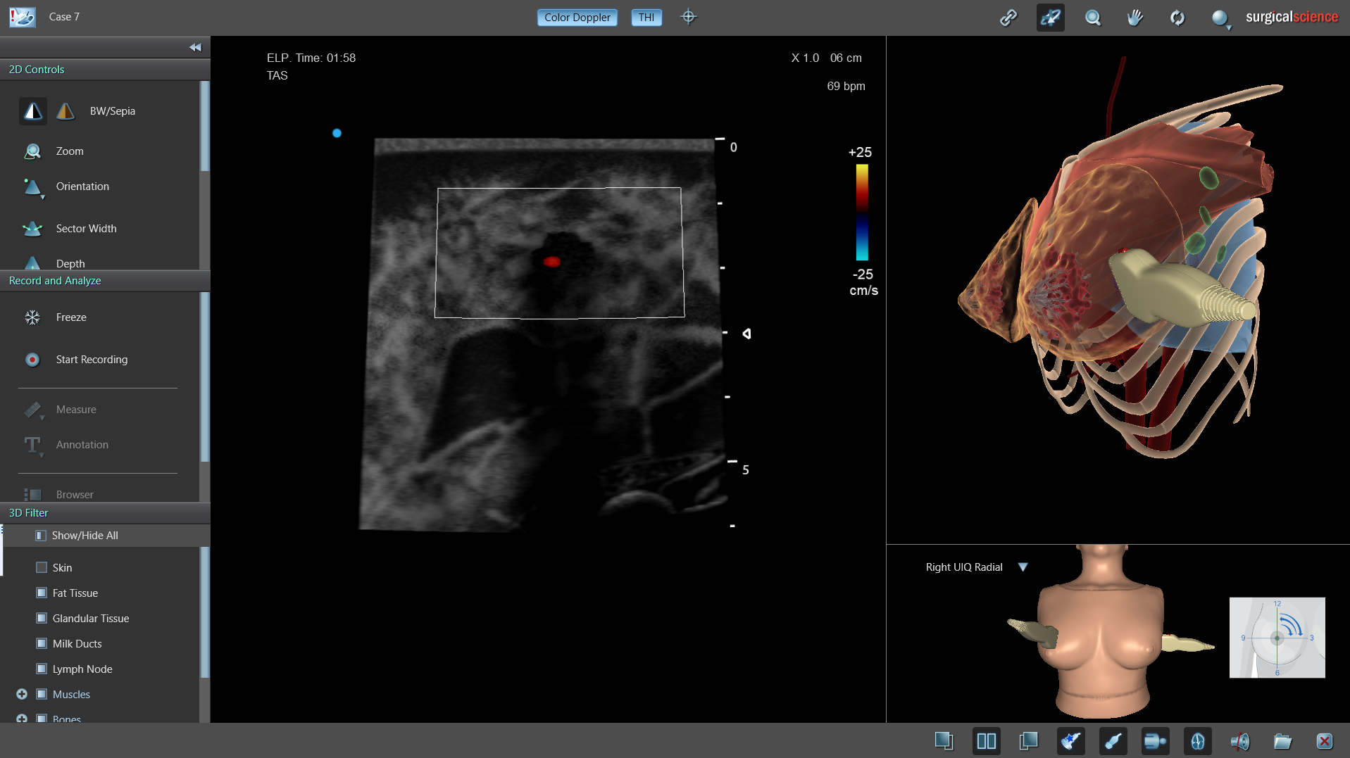
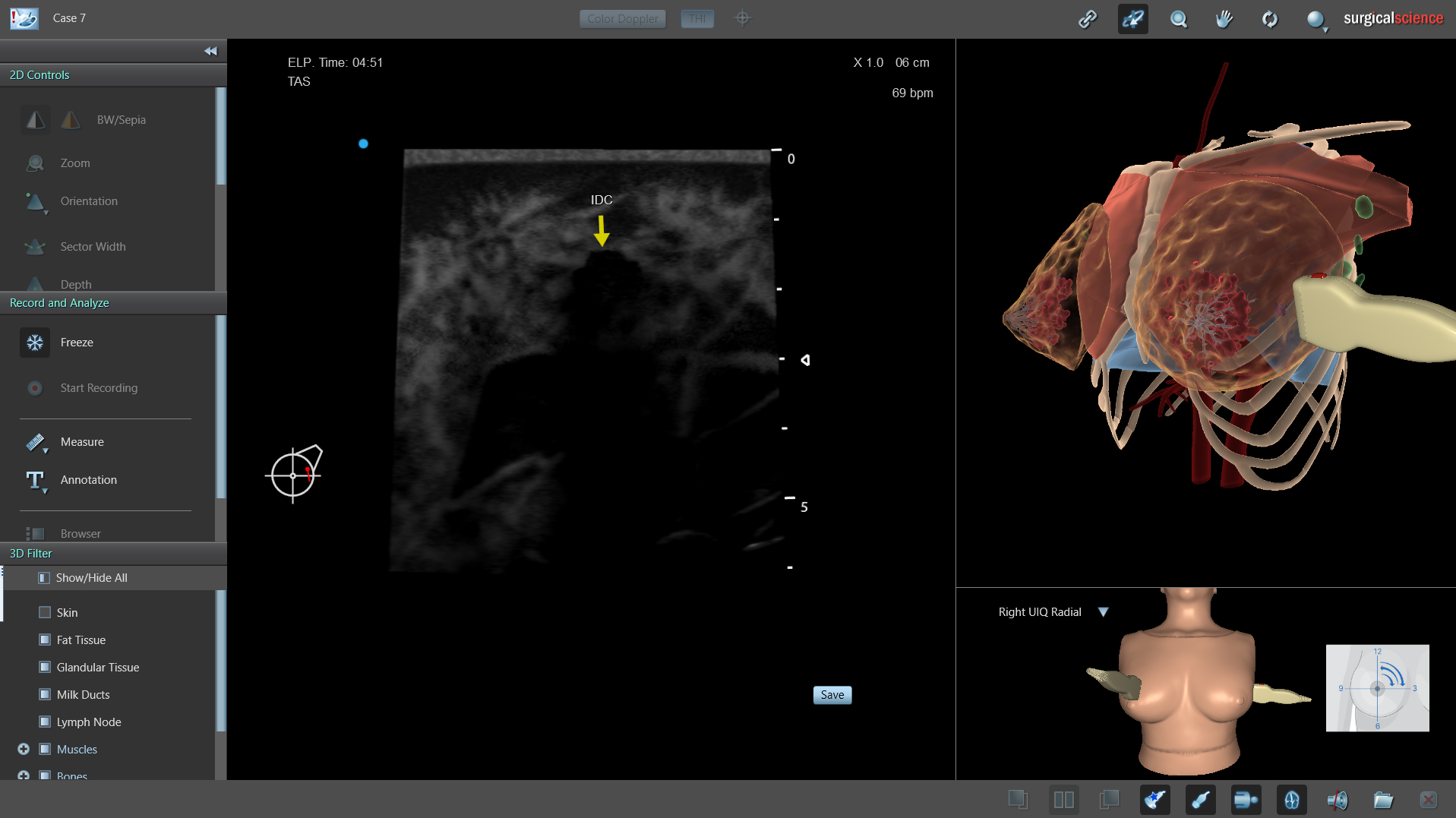
The Breast Ultrasound Module on Ultrasound Mentor on Ultrasound Mentor provides structured, hands-on training in breast ultrasound examination. Combining guided scanning tasks with simulated cases and cases based on recorded real ultrasound volumes, it enables learners to develop consistent scanning techniques without relying on live patient encounters.
A dedicated breast add-on provides a realistic scanning experience, including tissue compression and variations in breast composition. Learners can explore healthy anatomy and a range of pathologies, from simple cysts to invasive carcinoma. Repeatable examinations and objective performance metrics support feedback and competency assessment.
The module includes:
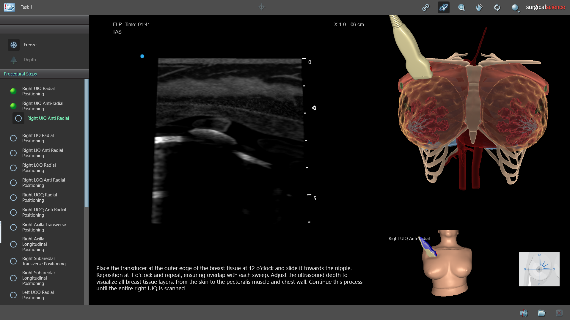
- Two guided scanning tasks: Step-by-step guidance focused on systematic scanning and coverage of the breast and axilla.
- Nine simulated cases: Cases covering healthy breast anatomy and a range of pathologies.
- Three cases based on recorded real ultrasound volumes: Opportunities to apply scanning and interpretation skills using recorded ultrasound imaging.
Additional module capabilities allow learners to practise image acquisition, annotation and documentation of findings.
Learning Objectives
- To practise systematic scanning and coverage of the breast and axilla.
- To develop radial/anti-radial and longitudinal/transverse breast scanning techniques.
- To identify breast anatomy and recognise variations in healthy breast composition.
- To practise probe positioning and tissue compression to obtain high-quality ultrasound images.
- To recognise and evaluate the sonographic appearances of varied breast pathologies.
- To apply scanning and interpretation skills using cases based on recorded real ultrasound volumes.
- To practise image acquisition, annotation and documentation of examination findings.
Hardware Requirements: The module requires a dedicated breast add-on for the Ultrasound Mentor female manikin.
Speak to Our Experts
Find out how the Breast Ultrasound module can expand your Ultrasound Mentor training capabilities and support your breast imaging curriculum.